
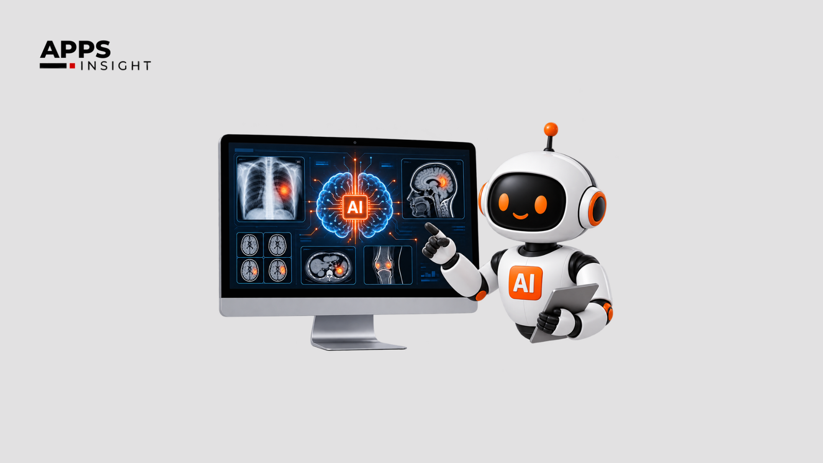
AI in Medical Imaging helps radiologists analyze scans, prioritize urgent cases, identify possible abnormalities, automate repetitive tasks, and improve workflow efficiency. It can examine X-rays, CT scans, MRI images, mammograms, and other studies for patterns that may require closer review. The technology is generally used as decision support rather than as a replacement for the radiologist. Evidence from mammography and other imaging studies shows that AI can reduce workload and, in some settings, improve sensitivity or reporting speed, although results depend on the specific tool, clinical setting, data quality, and level of human oversight.
For hospitals and healthcare businesses, the practical value is therefore less about replacing clinical expertise and more about helping radiologists spend their time on cases and decisions where human judgment is most important.
Related Queries
What are the main benefits of AI in medical imaging?
The main benefits include faster image analysis, case prioritization, support for abnormality detection, reduced repetitive workload, and more consistent workflow.
How does AI help radiologists?
AI can analyze medical images for predefined patterns, flag potentially urgent findings, organize worklists, and provide a second layer of analysis for the radiologist.
Can AI reduce radiologist workload?
Several studies have reported substantial workload reductions in specific imaging workflows, particularly breast cancer screening, although the actual reduction varies by use case.
Can AI improve diagnostic accuracy in radiology?
AI can improve sensitivity or specificity for selected findings, but performance varies by disease, imaging modality, patient population, and software. It should be evaluated for its intended clinical use rather than treated as universally accurate.
Will AI replace radiologists?
Current clinical use is primarily focused on assisting radiologists. AI can automate specific tasks, but radiologists remain responsible for interpreting findings in the patient’s clinical context and making appropriate decisions.
Why Are Radiologists Using AI?
Radiology is highly dependent on visual analysis. A radiologist may review hundreds of images in a single study and may need to compare them with previous examinations, identify subtle findings, prioritize urgent cases, and prepare a detailed report.
This creates a practical challenge: how can radiologists process growing imaging volumes without allowing speed to compromise careful review?
AI in Medical Imaging addresses part of this problem by using machine-learning and computer-vision models to analyze images and identify patterns. Depending on the application, an AI system can highlight a suspected lung nodule, flag a possible intracranial hemorrhage, classify a mammogram by risk, check image quality, or move potentially urgent examinations higher in a worklist.
The important point is that AI does not have to make the final diagnosis to be useful. Its value can come from handling specific, repetitive, or time-sensitive tasks while the radiologist remains responsible for clinical interpretation.
The technology is already part of the regulated medical-device landscape. The U.S. Food and Drug Administration maintains a list of AI-enabled medical devices authorized for marketing in the United States and notes that listed devices have gone through applicable premarket requirements for safety and effectiveness.
How Does AI Work in Medical Imaging?
At a basic level, an imaging AI system is trained to recognize patterns in medical images. For example, developers may train a model using a large collection of chest X-rays labeled according to whether particular abnormalities are present. During development, the model learns statistical patterns associated with those labels. When a new image is submitted, the software produces an output such as a probability score, alert, measurement, segmentation, or highlighted area. The output depends on the intended use. Common applications include:
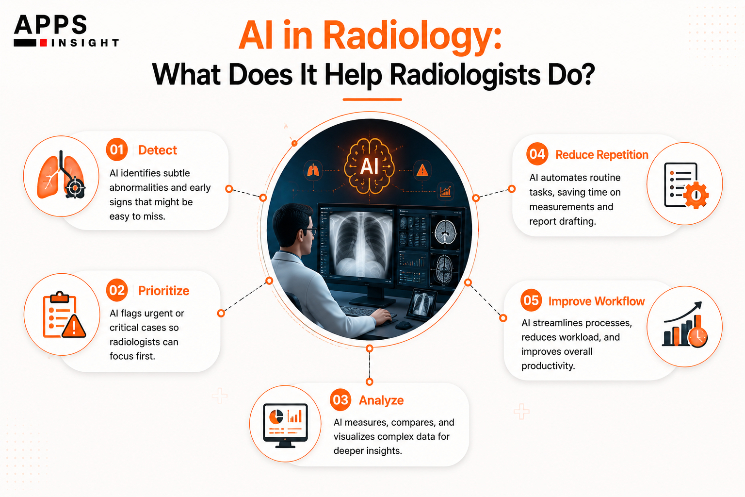
- Detection: identifying possible abnormalities such as nodules, fractures, or bleeding.
- Classification: assigning images or findings to categories.
- Triage: moving potentially urgent examinations higher in the radiologist’s worklist.
- Segmentation: outlining structures or lesions to help with measurement and analysis.
- Quantification: calculating measurements such as lesion size or organ volume.
- Image-quality assessment: identifying examinations that may be technically inadequate.
- Reporting support: assisting with structured information or repetitive documentation.
For example, a chest X-ray AI system may flag a suspected abnormality and present the result alongside the original image. The radiologist then reviews the image, considers the patient’s history and other evidence, and decides whether the finding is clinically meaningful.
A 2025 meta-analysis of 15 studies involving approximately 12,000 chest X-rays reported pooled AI sensitivity of 88% and specificity of 90% for pneumonia detection. For lung nodules, pooled sensitivity was about 72% and specificity about 95%. These figures show why performance must be considered by use case rather than assuming one accuracy number applies to every imaging task.
What Are the Benefits of AI in Medical Imaging for Radiologists?
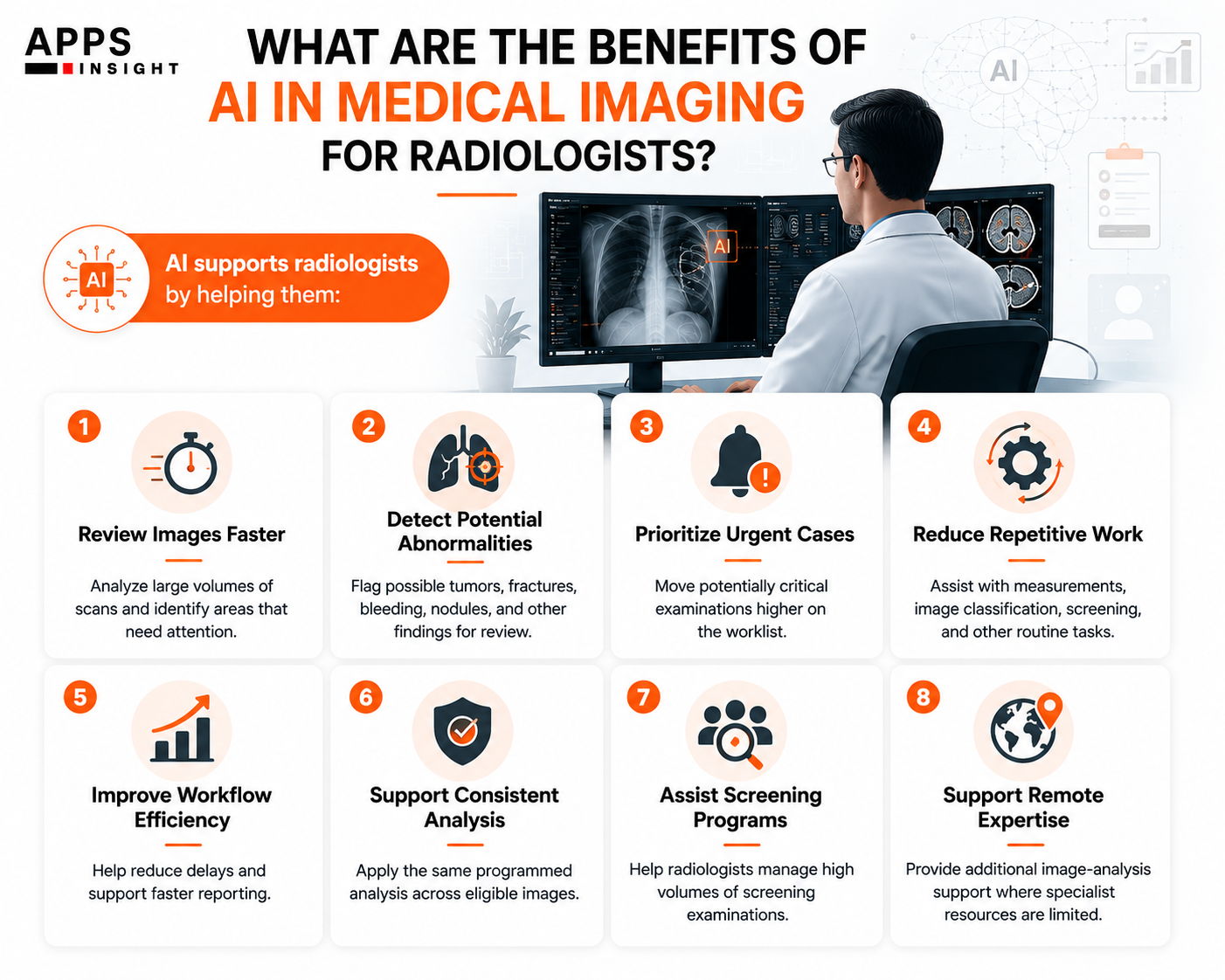
1. Faster review of large imaging volumes
One of the clearest benefits is the ability to process images quickly. AI can examine an image set in seconds and flag specific findings before or while a radiologist begins interpretation. This can be useful when departments handle high imaging volumes or when certain findings require rapid attention.
For example, in emergency radiology, an AI system may identify a possible intracranial hemorrhage and place the examination higher on the worklist. The radiologist still reviews the scan, but the system can help reduce the chance that an urgent case remains buried among routine examinations.
A 2025 systematic review of deep-learning worklist triage found reductions in report turnaround time across several clinical categories. The review reported mean differences of approximately 12.3 minutes for pulmonary embolism and 20.5 minutes for stroke, although the effect varied by setting and implementation.
This is particularly relevant for hospitals where turnaround time affects downstream care. Faster prioritization can help clinicians receive important imaging information sooner.
2. Reducing repetitive workload
Radiologists do not spend all of their time making complex clinical judgments. Some parts of the workflow involve repetitive image review, measurements, quality checks, and screening tasks. AI can assist with these activities.
For example, a breast-screening system can identify examinations that appear low risk and help prioritize those that need closer review. In a large retrospective simulation involving 114,421 mammography screenings, an AI-based screening protocol reduced the modeled radiologist workload by 62.6%. The study also reported AI sensitivity of 69.7%, compared with 70.8% for radiologist screening, while specificity was higher in the simulated AI protocol.
A 2024 meta-analysis of three studies involving 156,852 mammography examinations reported a theoretical radiologist workload reduction of 68.3% under the evaluated AI triage approach, with sensitivity of 93.1%. However, the researchers also noted substantial heterogeneity between studies.
These numbers should not be interpreted as meaning every radiology department can remove two-thirds of its workload. They demonstrate that AI can reduce workload for specific tasks when the technology, threshold, workflow, and clinical population are appropriate.
3. Supporting detection of subtle findings
Another important benefit is acting as an additional set of computational eyes. Radiologists already use clinical history, previous images, measurements, and their own experience when interpreting studies. AI can add another layer of analysis by looking for predefined image patterns.
This may be useful for findings that are small, subtle, numerous, or easy to overlook during a busy shift.
For example, an AI tool may highlight a possible pulmonary nodule on a CT scan. The radiologist can then examine the highlighted area and decide whether it represents a real finding, an artifact, or a clinically insignificant feature.
Research in chest trauma provides another example. A 2026 systematic review and meta-analysis found that AI assistance for rib-fracture detection was associated with increased diagnostic sensitivity and a reduction in detection time. However, the authors also rated the overall evidence quality as poor and called for further research.
That qualification matters. AI should be treated as an aid whose performance must be tested in the environment where it will actually be used.
4. Improving workflow consistency
Radiology departments need consistent processes, especially when imaging volumes are high. AI can apply the same programmed analysis to every eligible examination. This can help with tasks such as image-quality assessment, prioritization, measurements, and structured classification.
For instance, an image-quality tool can check whether an examination meets certain technical requirements before it reaches the reporting stage. If the image is inadequate, staff may be able to address the problem earlier.
A 2026 study evaluating an AI image-quality module integrated into a PACS/worklist reported lower median image-quality assessment time and improved report turnaround time after implementation. The study also found improved sensitivity for identifying suboptimal CT and MRI examinations without reducing specificity.
For hospital leaders, this illustrates an important point: useful AI does not always have to diagnose disease. Improving the quality and flow of information around diagnosis can also create operational value.
5. Helping radiologists prioritize urgent cases
Not every imaging examination has the same clinical urgency. A patient with suspected stroke, internal bleeding, or another time-sensitive condition may require faster review than a routine follow-up examination.
AI-supported triage can analyze incoming studies and identify cases that meet predefined criteria. These cases can then be moved higher in the worklist.
This approach can be particularly useful in large hospitals where imaging studies arrive continuously from emergency departments, inpatient units, outpatient clinics, and specialist services.
The benefit is not simply “faster AI.” The real benefit is better allocation of radiologist attention. A radiologist can spend less time deciding which case to open next and more time interpreting the studies that require immediate attention.
6. Supporting screening programs
Screening programs can generate very large numbers of examinations, making efficiency especially important. Mammography is one of the most studied examples. A 2024 population-based study comparing periods before and after AI implementation reported a 33.5% reduction in reading workload. The study also reported a lower recall rate, from 3.09% to 2.46%, and an increase in cancer detection rate from 0.70% to 0.82%.
A more recent prospective trial involving 31,301 women reported a 63.6% reduction in radiologist workload under the evaluated AI-supported screening strategy and a 15.2% higher cancer detection rate. However, the recall rate was 14.8% higher, showing that benefits can come with trade-offs that need clinical evaluation.
This is why the Benefits of AI in Medical Imaging should be evaluated using several measures together, including sensitivity, specificity, recall rate, false positives, turnaround time, workload, and patient outcomes.
7. Making imaging expertise more accessible
AI can also support radiology services where specialist availability is limited. A smaller hospital, rural facility, or imaging center may not have immediate access to a subspecialist for every type of examination. AI may provide preliminary analysis or prioritization while the final interpretation remains with a qualified clinician.
This does not eliminate the need for radiologists. Instead, it can provide additional support when workloads are high or specialist resources are unevenly distributed.
The World Health Organization recognizes AI as an existing technology in areas such as diagnosis and clinical care, including radiology and medical imaging. At the same time, WHO emphasizes safety, ethics, accountability, transparency, and human control when AI is used in healthcare.
How Does AI Fit Into a Radiology Workflow?
A typical AI-supported workflow can be relatively simple. First, an imaging study is acquired through an X-ray, CT, MRI, mammography, ultrasound, or another modality. The images are stored in the hospital’s imaging infrastructure, commonly through PACS.
The AI system receives the appropriate image data and performs its specific analysis. It may return an alert, probability score, measurement, annotation, or classification. The result is then presented to the radiologist through an appropriate clinical interface.
The radiologist reviews both the original images and the AI output. If the AI has flagged a suspected abnormality, the radiologist determines whether the finding is valid and clinically relevant.
The final report remains part of the clinical workflow. Integration is therefore just as important as model performance. A highly accurate algorithm that creates extra clicks, interrupts the radiologist’s workflow, or produces too many unnecessary alerts may provide limited practical value.
For organizations planning implementation, Hospital Management Software may also need to connect with imaging systems, patient records, scheduling, billing, and other operational systems so information can move through the wider hospital workflow.
Similarly, AI solution providers should be evaluated not only on model performance but also on interoperability, security, regulatory status, monitoring, support, and clinical validation.
What Should Hospitals Consider Before Adopting AI?
AI should be introduced for a clearly defined problem rather than simply because the technology is available. Hospitals should first identify the workflow problem. Is the goal to reduce reporting delays? Prioritize emergency cases? Improve screening? Reduce repetitive measurements? Improve image quality? The answer determines what type of AI is appropriate.
The next step is evidence evaluation. Organizations should ask:
- What clinical problem does the model solve?
- What population was used to validate it?
- Which imaging modalities and devices were included?
- What are its sensitivity and specificity?
- How often does it produce false positives?
- Has it been evaluated in a real clinical workflow?
- Does it integrate with existing PACS and clinical systems?
- How is patient data protected?
- How are model updates evaluated?
- What happens when the AI system is unavailable?
- Who remains responsible for the final clinical decision?
AI in healthcare solutions also needs appropriate governance because medical data is sensitive and clinical decisions can have serious consequences.
WHO’s guidance on AI in healthcare identifies principles including protecting human autonomy, promoting safety, ensuring transparency and explainability, establishing accountability, supporting equity, and developing AI that is responsive and sustainable.
Organizations evaluating vendors can also use independent technology directories and research platforms. AppsInsight provides listings covering healthcare technology, AI, software development, and related IT services. Its healthcare-focused listings can be useful as one source for building an initial vendor shortlist, although hospitals should conduct their own technical, clinical, security, and regulatory due diligence before selecting a provider.
For teams building connected healthcare products, Healthcare Software Development Companies may also be relevant when AI needs to work with EHRs, PACS, patient applications, or other clinical systems.
EHR Software Development Companies can help organizations connect imaging results with patient records and broader clinical workflows.
In some care models, Telemedicine Software Development Companies can also become part of the wider architecture when imaging needs to be reviewed remotely by specialists.
Telehealth Software Development Firms may similarly support remote consultation and access to imaging data, especially for organizations using distributed care models. The key is to treat AI as one component of a clinical system rather than as a standalone product.
What Are the Limitations of AI in Medical Imaging?
Before adopting AI for medical imaging, hospitals and radiology departments need to look beyond accuracy claims. You have to understand how the system performs in real clinical conditions, how it fits into existing workflows, and what risks it may introduce. A well-planned evaluation can help organizations choose technology that supports radiologists without creating additional workload or safety concerns.
1) Clinical Accuracy Can Vary
AI performance is not the same for every imaging study, disease, patient group, or hospital. A model trained and validated using one dataset may perform differently when it is used with images from another hospital, scanner, population, or clinical environment.
For example, an AI system may perform well when detecting a particular abnormality in chest X-rays but may be less reliable when images have different acquisition settings or when patients have conditions that were underrepresented in the training data.
Hospitals should therefore examine more than a single accuracy percentage. Important measures include sensitivity, specificity, false-positive rate, false-negative rate, and positive predictive value.
The technology should also be evaluated using data that reflects the population in which it will actually be used.
2) AI Can Produce False Positives and False Negatives
No medical AI system is perfect. An AI tool may flag an area as suspicious when it is normal, creating a false positive. It can also fail to identify a real abnormality, resulting in a false negative.
False positives can increase the number of cases that radiologists need to review, while false negatives can create patient-safety concerns if an important finding is missed.
This means AI output should generally be treated as decision support rather than an automatic diagnosis. The radiologist should review the original images, consider the patient’s clinical history, and make the final interpretation.
For healthcare organizations, understanding the error profile of a system is often more useful than looking at its overall accuracy alone.
3) Integration With Existing Systems Is Important
An AI tool may work well on its own but still provide limited value if it does not integrate smoothly with the hospital’s existing technology.
Radiology departments commonly use systems such as PACS, RIS, EHR platforms, imaging modalities, and reporting applications. AI needs to exchange information with these systems without creating unnecessary manual steps.
For example, if a radiologist has to leave the PACS, open a separate application, upload an image, wait for the result, and then manually transfer the information back into the reporting workflow, the technology may add friction instead of reducing it.
Before implementation, hospitals should check:
- PACS and RIS compatibility
- Data exchange standards
- EHR integration
- Workflow and worklist integration
- Processing time
- User-interface requirements
- System availability and downtime procedures
Good integration allows AI results to appear where radiologists already work.
4) Data Privacy and Security Must Be Addressed
Medical images contain sensitive patient information. Hospitals need to understand how imaging data is collected, processed, stored, transferred, and deleted when using an AI system.
Organizations should ask vendors whether patient information is processed locally or through cloud infrastructure and what security controls are used to protect the data. Important questions include:
- Where is patient data stored?
- Who can access it?
- Is data encrypted during transfer and storage?
- Is patient information used to train future models?
- How long is the information retained?
- What happens to the data when the contract ends?
- How are security incidents handled?
These questions become especially important when AI platforms connect with multiple clinical systems or external services.
5) Regulatory Approval and Intended Use Should Be Checked
Healthcare organizations should verify whether an AI product has the appropriate regulatory authorization for its intended use and market.
Regulatory status does not mean that an AI system is suitable for every clinical purpose. A product may be authorized for one specific indication, imaging modality, or clinical workflow but not for unrelated uses.
The U.S. Food and Drug Administration maintains a public list of AI-enabled medical devices authorized for marketing in the United States. The FDA also emphasizes that these devices are subject to applicable regulatory requirements.
Hospitals should therefore review the product’s intended use, regulatory documentation, clinical evidence, and applicable requirements before deployment.
6) AI Can Create Additional Alerts
One common concern is alert fatigue. If an AI system produces too many notifications, radiologists may start receiving a large number of alerts that do not require immediate action. Instead of simplifying the workflow, excessive alerts can make it harder to identify genuinely urgent cases.
For example, an AI system designed to flag possible abnormalities may generate several alerts during a busy shift. If many of those findings are ultimately considered normal, the radiologist has to spend additional time checking them.
Organizations should evaluate the alert threshold before full deployment and monitor how often AI-generated alerts result in clinically meaningful findings.
7) Staff Training Is Still Necessary
Introducing AI does not remove the need for professional training. Radiologists and other clinical staff need to understand what the system is designed to detect, how its results are displayed, and what its limitations are. They should also know what to do when the AI result conflicts with their own interpretation. Training should cover practical questions such as:
- What does an AI score mean?
- When should an alert be reviewed?
- What findings can the system miss?
- How should incorrect results be reported?
- What should staff do if the system is unavailable?
- Who is responsible for the final clinical decision?
The goal is not to train radiologists to blindly follow AI recommendations. It is to help them use the system appropriately as an additional source of information.
8) Performance Needs Continuous Monitoring
AI performance should not be treated as a one-time evaluation. After deployment, hospitals should continue monitoring whether the system performs as expected. Changes in imaging equipment, patient populations, clinical workflows, or software versions can affect performance. A useful monitoring program can track:
- Detection performance
- False-positive and false-negative rates
- Radiologist workload
- Report turnaround time
- Number of overridden AI recommendations
- Alert volume
- System downtime
- User feedback
- Patient and clinical outcomes where appropriate
Continuous monitoring can help identify problems before they become widespread.
9. Cost Should Be Evaluated Against Practical Value
The cost of AI adoption includes more than the software subscription or licensing fee. Hospitals may also need to pay for integration, infrastructure, cybersecurity, training, technical support, maintenance, and ongoing monitoring.
For this reason, organizations should define the expected outcome before purchasing a solution. For example, if the main goal is reducing emergency report turnaround time, the hospital can measure turnaround time before and after implementation. If the goal is reducing repetitive screening workload, it can measure reading time and case volume.
This creates a clearer way to determine whether the technology is providing meaningful operational value.
10. Human Oversight Should Remain Central
The most important consideration is the role of the radiologist. AI can process images quickly and identify patterns, but clinical interpretation involves more than recognizing a visual feature. Radiologists consider symptoms, medical history, previous examinations, laboratory results, treatment history, and other clinical information.
An AI system may identify a suspicious lesion, for example, but the radiologist needs to determine what that finding means in the context of the patient.
The World Health Organization emphasizes human autonomy, safety, transparency, accountability, and appropriate governance when AI is used in healthcare. For this reason, healthcare organizations should design AI-supported workflows where clinicians can review, question, and override AI results when necessary.
We have listed! Profitable Healthcare Business Ideas
Final Say
AI can provide practical support to radiologists by analyzing images, identifying possible abnormalities, prioritizing urgent examinations, reducing repetitive tasks, supporting screening, and improving parts of the radiology workflow.
The strongest evidence is not that AI universally makes radiologists faster or more accurate. Instead, research shows that specific AI applications can produce measurable benefits for specific tasks and clinical settings. Mammography studies, for example, have reported substantial reductions in reading workload, while other studies have shown improvements in turnaround time or detection sensitivity.
AI in Medical Imaging is therefore best understood as a clinical support technology. Its value depends on the problem being solved, the quality of the model, the patient population, workflow integration, and the ability of clinicians to review and act on its output.
For radiologists, the most useful systems are likely to be those that remove repetitive work, surface important cases, and provide meaningful information without creating additional noise.
For hospitals and technology buyers, the right question is not simply whether AI is available. It is whether a particular AI system can solve a clearly defined clinical or operational problem, produce reliable results, integrate into existing systems, and do so with appropriate safety and human oversight.
When those conditions are met, AI in Medical Imaging can become a practical part of modern radiology rather than another layer of technology added to an already complex workflow.

Sources and Further Reading
- U.S. FDA: Artificial Intelligence-Enabled Medical Devices
- PubMed: AI-based mammography screening and radiologist workload
- PubMed: AI triaging of breast cancer screening mammograms, meta-analysis
- PubMed: AI impact on mammography screening performance and workload
- PubMed: AI-based mammography noninferiority trial
- PubMed: AI diagnostic accuracy in chest radiography
- PubMed: Deep-learning worklist triage and radiology turnaround time
- World Health Organization: Ethics and governance of AI for health
- World Health Organization: Ethics and medical radiological imaging
- AppsInsight: Healthcare and technology company directory